
register online
pay online
|
 |
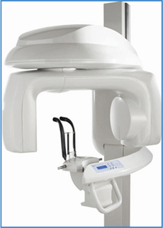
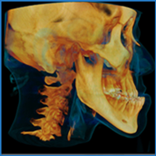
3-Dimensional Imaging and Cone Beam Computed Tomography (CBCT) Cone Beam Computed Tomography (CBCT) is a 3-Dimensional imaging modality providing the best possible maxillofacial imaging techniques available today.
CBCT allows us to obtain images of the skull and jawbones in a relatively short period of time (approximately 10-15 seconds). This information can then be used to evaluate you in a way that was previously only possible with expensive medical CT scans. Perhaps the most compelling example is implantology. In order to perform a successful implant, the dental professional must have a precise understanding of the structure of the jawbone. This understanding ensures that the implant is the optimum width and length to provide the necessary strength while maintaining the integrity of the bone in which it is placed. Three-dimensional imaging exams are ideally suited to providing all the information needed to choose the appropriate implant and position it accurately. The data generated by many 3D dental imaging systems can be used to create a surgical drilling guide to facilitate rapid and accurate placement of implants; it can also be used to create custom treatment appliances. Post-operatively, three-dimensional imaging exams can be used to verify that an implant was properly aligned. While we do not obtain a CBCT on every patient, we do evaluate each patient on an individual basis to determine the benefits that may be obtained with a CBCT. |
  |
What is CBCT used for?
What are the advantages of CBCT over other imaging modalities?
|
 Members of the American Association of Oral and Maxillofacial Surgeons Members of the American Association of Oral and Maxillofacial Surgeons |
 Diplomates of the American Board
of Oral and Maxillofacial Surgery Diplomates of the American Board
of Oral and Maxillofacial Surgery
|
| * * Board Eligible * Emeritus |
|
| Oral Surgery Website: |
| Dental Website Design By omswebsites.com. - Copyright ©2011 |


